Editor’s NOTE: The reference to a ”100% cure rate in Terminal Panceatic Malignancies‘?is based upon the 1986 Memorial Sloan Kettering review of Dr. Kelley’s Pancreatic Cases, conducted by Dr. Nicholas Gonzalez.
Kathy P. Fairbanks, Ph.D.: What is Cancer: “Cancer is a process misunderstood by the medical community. Cancer is classified by the medical community, as a fast-growing malignant tumor, which, if allowed to grow unchecked, will cause death.”
 Many clinicians believe that cancer is a complex: a number of different diseases, each having it’s own cause. Most doctors, even research scientists, suppose such things as viruses, X-rays cigarette smoking chemicals, sunlight, and trauma causes cancer. However there are a growing number of cancer researchers who believe that these factors, rather than causing cancer, are indirect stimulators of a normal trophoblast-like pleuripotential cell. This trophoblast-like cell then makes its “faIse placenta”, a malignant tumor mass, which the medical community calls cancer.
Professional Review and Update: Chapter I (Revised)
~ In the Beginning ~
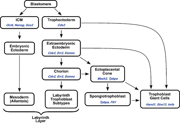
In the first five days after fertilization in the formation of a human embryo, the growing mass of cells divides into two kinds of cells, an inner cell mass (embryoblasts) which become the embryo, and an outer layer of cells called the trophoblast, which later forms the placenta. This process is so complex that half of the developing masses ever progress past this stage. Something goes wrong with normal development and they are expelled from the woman’s body before they can implant themselves in the uterus.
After the cell mass attaches to the wall of the uterus, the trophoblasts invade the lining of the uterus, growing quickly and invasively, as a tumor does when invading an organ of a human body. The trophoblast cells invade, digest a bole in the wall of the uterus and form a multinucleated mass with no cell boundaries, which looks under the microscope like the cells of a carcinoma. During this invasion of the trophoblasts into the uterine wall, the pregnant woman may feel nauseous with “morning sickness” due to the trauma of being assailed by this cancer-like mass. As small blood vessels are invaded and digested by the invading trophoblasts, pools of blood form in the tissue, which nourishes the growing mass. The failure of the maternal tissue to reject this implantation has always puzzled embryologists and immunologists. One current view is that the trophoblast cells lack certain protein on their surfaces, and thus are not recognized as foreign by the mother’s body.
~ Primary Germ Cell ~
During the time that the trophoblast cells are aggressively infiltrating the maternal tissue, the inner cell mass is organizing itself into a three part disc, shaped like I flying saucer. These three parts of the disc are called the three primary germ layers, or the ectoderm, the endoderm and the mesoderm. Each of these three layers become different parts of the human body. The ectoderm becomes the skin, the brain and the nerves. “Ecto” means surface, and indeed these cells become the surface covering of the body, and the nerves which are the interface, of the body with the outside world. The endoderm becomes the linings of many organs) such as the lungs, the intestines, liver, and pancreas “Endo” means within, and indeed these cells become almost all of the linings of the body. The mesoderm becomes the muscles, blood, bone, and the reproductive organs. “Meso” means middle, and these mesoderm cells, which form as the middle layer of the disc, become the vast majority of the cells of the body, almost all of the different cell types.
 This process of organ formation involves extensive migration of certain cells from the disc to their future sites. The mesoderm cells come from an area on the disc known as the primitive streak. Under a microscope, a dark streak progresses visibly along the center of the disc from the tail end to the head end of the disc. This primitive streak is caused as ectodermal cells drive down into the middle of the disc like the filling of a sandwich, becoming mesodermal cells in the process. This migrating of ectodermal cells becoming mesodermal cells happens very early in development – between two weeks and three weeks after the trophoblasts begin invading the uterus of the mother. These migrating cells, which come from the primitive streak, are pleuripotent. The mesoderm cells are called pleurlpotential, because under different circumstances they are able to follow more than one pathway of development. In other words, mesoderm cells can potentially form many kinds of tissue. They are cells, which are closest in nature to the unruly aggressive trophoblastic cells that have formed the placenta.
This broad developmental potential of the pleuripotential cells becomes more and more restricted and checked as the tissues acquire the specialized control mechanisms to guide the cells in their development. Increasingly complicated migrations of cells occur as the body of the new human is forming. For instance in the ectoderm, neural cells migrate in the myriad directions and become specialized neurons. This regimentation of a cell’s capabilities must occur in order to form, for example, a bone cell as opposed to a muscle cell in the mesoderm. Such regimentation comes about in response to cues from the immediate surroundings, including the nearby tissue. The precision and coordination required for correct development is dependent upon these interactions. Thus, nearby tissues influence development of certain cells, probably by signals carried by certain protein molecules. Interestingly enough, these signals must also occur at a certain precise time, so that a delay in this signal may lead to the failure of correct interaction, leading to various kinds of defects. Many of these defects cause the death of the developing embryo, and some lead to birth defects.
~ Direct Cause of Cancer ~
 The intricate and precise orchestration of the formation of a normal human from the original inner cell mass is a miracle of precision timing and maturation of these pleuripotential cells. Every normal human contains varying numbers of cells, which have not completed their correct migrations, thereby leaving “sleeping” pleuripotential cells scattered throughout the body. When these pleuripotential cells are activated through genetic, environmental or nutritional factors, a tumor cell mass, similar to the invasive trophoblastic cell mass can begin to form. This cancerous tumor may contain various types of tissue, such as chips of bone or hair. These scattered pleuripotential cells are normally prevented from becoming a cancerous tumor through circulating protein molecules, which keep their growth in check. It has been theorized that when a human body does not have enough of these patrolling molecules, the pleuripotential cell grows in an unrestrained fashion, becoming a carcinoma.
In summary, the early embryo has two cell types: the trophoblast and the embryoblast. The embryoblast becomes the three germ cell types; the ectoderm, the endoderm and the mesoderm. The mesodermal cells are pleuripotential, with a vast ability to become many different kinds of cells. Some of these remain “sleeping” dispersed throughout the tissues of the body.
~ How Do Enzymes Work? ~
 Enzymes are normally produced by the pancreas to help digest the food that enters the small intestine from the stomach. Different kinds of enzymes work on protein, on fats or on starch and sugar. By the action of these powerful enzymes, large particles of protein, fat or starch, are broken down into smaller and smaller pieces, until they are small enough to go through the wall of the small intestine and be used there to digest food coming back from the stomach. These enzymes can also be absorbed through the wall of the small intestine into the body, and travel in the blood stream to distant locations in the body where they are needed.
Why don’t these powerful enzymes start dissolving the very tissue that they are passing through? How can these enzymes travel to the tumor and only digest the cancer, without harming the person’s body in which the cancer is growing? The secret to how the enzyme can tell the difference between “good tissues and bad tissues”, lies in a difference as small as the difference between your right hand and your left hand. Almost all the billions of tiny molecules in the body are either right-handed or left-handed. As an example of right and left handedness, let’s look at a pair of mittens. In a pair of mittens you find one for the right hand and one for the left hand. They are mirror images of each other, but if you tried to put the right-handed mitten down on top of the left-handed mitten, they would not match. In a mysterious way, the human body uses only right handed sugar molecules, but only left-handed protein molecules.
The above paragraph has discussed right-handed sugar molecules and left-handed protein molecules. Logic raises the question, where are the mirror image substances? Where are the left-handed sugar molecules and the right-handed protein molecules? These are found within the placenta, which is made of trophoblasts. They are also found within the trophoblast-like tumor cells. What difference does this make for the enzyme trypsin?
We know that the enzyme trypsin acts on cooked left-handed proteins and living (non-cooked) rlght-handed proteins. Normally, when we eat a meal, the cooked left-handed proteins, which we eat, are digested in the small intestine by the trypsin released by the pancreas. Trypsin does not act on the organs of the human body, because these are living, left-handed protein. However, trypsin is very effective at breaking down living, right-handed proteins. And where did we say living right-handed proteins could be found? These living, right-handed proteins are the substance comprising the cancerous tumor. So, the trypsin can travel via the bloodstream to the tumor, and its action there is on the protein mass that makes up the tumor. It breaks down the protein mass of the tumor and “liquefies” it.
As further explanation, this cancerous tumor needs an enzyme with which it can digest the organ or tissue of the human where the tumor is located. It uses human tissue as food. To obtain its needed enzyme, the tumor itself makes the enzyme. This tumor-made enzyme is called malignin, which does digest human protein. This malignin is the mirror image enzyme to trypsin. In other words, trypsin and malignin are mirror images of each other, as your right hand and left hand are mirror images of each other. As trypsin acts on living right-handed protein, namely the tumor mass, so malignin acts only on living left-handed proteins, namely human tissue.
Trypsin in sufficient quantities can begin to break down the cancerous tumor but not fully digest the cancerous tumor. During the breakdown process, trypsin produces some intermediate proteins, which can be quite toxic to the human body. These intermediate proteins need a second enzyme to complete their digestion, i.e. “liquefaction”. Therefore, to be successful, the enzyme treatment for cancerous tumors must include both of these enzymes in sufficient quantities to render the products of tumor digestion harmless.
These enzymes work by traveling through the bloodstream to the site of the tumor and digesting the specific protein of the tumor mass, without harming the body’s tissues at all. This fascinating story of the matching rlght and left handed molecules, trypsin and malignin, was explained almost a century ago by Scottish doctor, John Beard, D.Sc. His revolutionary book, published In London in 1911, was entitled, The Enzyme Treatment of Cancer and Its Scientific Basis. At that time, some cancers were treated by direct injection of the enzymes near the cancer mass. Now, we realize that injecting the enzymes is unnecessary, since swallowing capsules containing the enzymes will also work. Trypsin will only digest the protein of the tumor, thus it can safely travel through the body.
The ability to target the tumor in such a specific and successful manner makes the use of surgery, chemotherapy and radiation obsolete.
Kathy P. Fairbanks, Ph.D.
~ Notes from Dr. Kelley ~
Professor Fairbanks’ scientific presentation above is pure truth and scientific without error. Any clinician who challenges this should start all over with his education – in high school biology.
I first read Dr. Beard’s book in May 2000. By 1962 I had developed my successful protocol and was free of cancer. I accept Professor Fairbanks’ most significant contribution to the understanding of my program, which should help the clinicians who stand up for proper treatment of those suffering with cancer.
The missing factors in Dr. Beard’s and Professor Fairbanks’ understanding are the enzyme activators, which have made my program so successful for the past 40 years. It has been my experience that without the complete program, success will be limited to approximately 52%.
Respectfully,
William D. Kelley, D.D.S., M.S.
Medical Missionary
